

")


Acta Universitatis Dentistriae et Chirurgiae Maxillofacialis
Peer-review medical journal.
Editor-in-chief
- Prof. Roman Fadeev, MD, PhD
Publisher
- Eco-Vector (Saint Petersburg, Russia)
Journal founders
- North-Western State Medical University named after I. I. Mechnikov
WEB: https://szgmu.ru/eng/ - Eco-vector Publishing Group (Saint Petersburg, Russia)
WEB: https://eco-vector.com/
About
The journal accepts manuscripts on the most significant issues of therapeutic, preventive and clinical studies in the field of dentistry, maxillofacial surgery, radiation diagnostics. When considering the received author's materials, the Journal is guided by the "ICMJE Recommendations". The Journal publishes previously unpublished works corresponding to the Journal profile. Multiple and duplicate publications are not allowed. Articles representing separate stages of incomplete research, as well as articles in violation of the Rules and Norms of Humane Treatment of Biological Objects of Research are not accepted for publication. Publication is possible only after receiving a positive review.
Sections:
- Reviews;
- Clinical dentistry and maxillofacial surgery;
- Scientific research;
- Publications of young scientists;
- Information about scientific conferences.
Publications
- quarterly, 4 issues per year
- russian and english articles and full-texts
Distribution
- Open Access, under the Creative Commons Attribution-NonCommercial-NoDerivatives 4.0 International License (CC BY-NC-ND 4.0)
Current Issue
Vol 3, No 2 (2025)
- Year: 2025
- Published: 15.09.2025
- Articles: 6
- URL: https://stomuniver.ru/unistom/issue/view/12971
- DOI: https://doi.org/10.17816/UDS.32

Clinical dentistry and maxillofacial surgery
Herpesvirus infection in dental patients
Abstract
BACKGROUND: Herpesviruses can persist asymptomatically, affecting the mucous membranes and skin across all age groups while remaining latent in neurons throughout life. Approximately 80% of patients exhibit no symptoms during lytic infection, resulting in missed treatment opportunities and inadvertent transmission to others, which facilitates the virus’s spread.
AIM: This work aimed to assess the prevalence of latent herpesvirus infections and their clinical manifestations in the oral cavity among dental patients.
METHODS: A total of 38 patients aged 10–17 years and 36 patients aged 19–35 years were examined. All patients underwent real-time polymerase chain reaction testing of saliva samples for the presence of Herpes Simplex Virus 1, Herpes Simplex Virus 2, Epstein–Barr Virus, Cytomegalovirus, and Human Herpesvirus 6. Results were compared between patients with positive and negative test outcomes, regardless of the specific virus identified.
RESULTS: Among patients aged 10–17 years, the following viruses were detected: Herpes Simplex Virus 1 in 27 patients, Herpes Simplex Virus 2 in 9, Epstein–Barr Virus in 12, Cytomegalovirus in 8, and Human Herpes Virus 6 in 18. In the 19–35-year-old group, Herpes Simplex Virus 1 was found in 12 patients, Herpes Simplex Virus 2 in 8, Epstein–Barr Virus in 18, Cytomegalovirus in 6, and Human Herpes Virus 6 in 19.
CONCLUSION: A high prevalence of asymptomatic Herpes Simplex Virus 1 infection (71%) was observed in patients aged 10–17 years, despite the absence of classical clinical signs. Among those aged 19–35 years, Human Herpes Virus 6 was most prevalent (53%).
 47-52
47-52


Clinical and functional parameters of dental status in children with cerebral palsy
Abstract
Background. High prevalence of dento-mandibular-facial anomalies, orofacial dysfunctions and accompanying complications in children with infantile cerebral palsy determine the urgency and significance of the problem of early detection and prevention of these types of pathology.
AIM. To estimate the frequency of orofacial dysfunctions and caries intensity in children with cerebral palsy.
Materials and methods. We examined 36 children with infantile cerebral palsy. The age ranged from 8 ± 5.2 years.
Results. It was revealed that 70% of the patients had mastication and swallowing disorders with severity up to 8.26±2.12 points. Up to 80% of patients during the daytime have oral type of breathing with a severity of 8.23±1.27 points. Also, 75% of patients have narrowing of the upper jaw up to 6.48±1.65 points and close position of teeth in frontal part of the upper and lower jaw. 27.8% of patients have low activity of the circular muscle of the mouth and the severity of the open bite was 5.23±1.23 points. Distal bite was detected in 66.7% of children. Crossbite is noted in 5.6%. Up to 90% of patients have asymmetry of masticatory muscles with the expression of 7.15±1.09. The prevalence of caries is 52% with an expression of 1.33±1.02
Conclusion. All children with cerebral palsy have orofacial dysfunction, asymmetry of masticatory muscles, and bite anomalies. The risk of dental caries development in this group of children is not high, because parents perform oral hygiene.
53-58
Scientific research
Occlusal–condylar circle on lateral cephalometric radiographs and its diagnostic value in clinical dentistry
Abstract
BACKGROUND: Advances in radiographic techniques continue to improve diagnostic capabilities, underscoring the relevance of this study.
AIM: This work aimed to determine the diagnostic value of the occlusal–condylar circle on lateral cephalometric radiographs in patients with different types of occlusal relationships.
METHODS: The study included two groups: group 1 comprised radiographs of patients with normal occlusion; group 2 included radiographs of patients with malocclusion and distal-extension edentulous defects. Geometric constructions were created on lateral cephalometric images using the PowerPoint software and established anatomical landmarks. The radius of the occlusal–condylar circle was defined as the distance from the superior point of the condylar head (Cond) to the distal occlusal point of the dental arch.
RESULTS: In group 1, the line of the occlusal–condylar circle passed through the distal occlusal point of the second molar and a point corresponding to two-thirds of the Cond–A line. In group 2, cephalometric analysis focused on occlusal point positions in cases of malocclusion and dental-arch defects due to missing molars with vertical dentoalveolar overeruption of antagonists.
CONCLUSION: The occlusal–condylar circle on lateral cephalometric radiographs can serve as a key reference for establishing the occlusal line in patients with malocclusion and in those who have molar-segment dental-arch defects. The findings may be applicable in clinical settings for diagnosing vertical occlusal anomalies and assessing the outcomes of prosthodontic treatment.
59-65
Impact of primary biliary cholangitis severity on oral health status
Abstract
BACKGROUND: Primary biliary cholangitis (PBC) is a chronic immune-mediated liver disease predominantly affecting women (prevalence: 2–40 cases per 100,000 population). It is often accompanied by xerostomia and marked oral changes. The pathogenetic mechanisms linking oral pathology and autoimmune liver disease remain poorly investigated.
AIM: This work aimed to correlate the pattern of oral changes in patients with PBC with disease duration, stage, and clinical-course characteristics.
METHODS: Forty-eight women aged 43 to 69 years with confirmed PBC were examined. Disease stage (early or advanced) was determined using laboratory data and transient elastography. Oral assessment included clinical dental examination, radiography, evaluation of salivary gland function (sialometry, salivary pH measurement, ultrasonography), and detection of key periodontopathogens via polymerase chain reaction. Statistical analysis was conducted using R software with regression modeling.
RESULTS: Advanced-stage PBC was identified in 52% of cases; xerostomia was present in 68.75%. Liver stiffness was positively correlated with xerostomia severity (τ = 0.54, p < 0.001), periodontitis stage (τ = 0.56, p < 0.001), and oral-hygiene status as well as the number of identified periodontopathogens. More severe oral abnormalities were observed with longer disease duration. Gamma-glutamyltransferase and alanine aminotransferase levels showed positive correlations with xerostomia and periodontitis severity.
CONCLUSION: Oral abnormalities in patients with PBC are closely associated with disease severity, duration, and liver function biomarkers. These changes may serve as supplementary clinical indicators of disease progression and support the need for a multidisciplinary approach to management.
67-72
Publications of young scientists
Effect of modified herbst appliance on the cervical spine in patients with skeletal class II malocclusion
Abstract
BACKGROUND: The potential impact of orthodontic treatment on the musculoskeletal system, particularly the cervical spine, remains a pertinent issue in orthodontics. Of special interest is the effect of functional appliances on cervical spine alignment in adult patients with skeletal class II malocclusion. The Herbst appliance is known to induce sagittal advancement of the mandible, which may affect postural balance, including the curvature of cervical lordosis.
AIM: This work aimed to evaluate cervical spine parameters in adult patients with skeletal class II malocclusion treated using a modified Herbst appliance.
METHODS: Cervical-spine morphology was analyzed on lateral cephalograms obtained from 15 adult patients with skeletal class II malocclusion who had been treated with a modified Herbst appliance. All radiographs were obtained using the Orthophos XG unit (Dentsply Sirona, Germany) in the natural head position.
RESULTS: Use of the modified Herbst appliance led to a mean reduction in cervical lordosis curvature of 1.36 ± 1.20 mm and a mean reduction in cervical lordosis angle of 3.00 ± 2.32.
CONCLUSION: The modified Herbst appliance may influence cervical spine alignment in adult patients with skeletal class II malocclusion. Further evaluation of postural adaptations, including pelvic and foot positioning before and after treatment, is needed to comprehensively assess treatment-related changes.
73-78
Information about scientific conferences
Legal framework of dentistry: summary of the first All-Russian interdisciplinary scientific and practical conference
Abstract
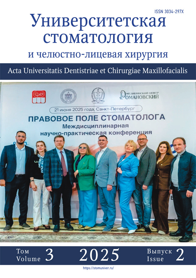
On June 21, 2025, the First All-Russian Interdisciplinary Scientific and Practical Conference titled “The Legal Framework of Dentistry” was held at the Grand Hotel Emerald conference hall in St. Petersburg. The event was organized by the North-Western State Medical University named after I.I. Mechnikov, in collaboration with the Russian Dental Association, the Scientific Medical Society of Dentists of St. Petersburg and the Leningrad Region, and the Romanovsky Medical Center. The event was attended by specialists in medical law and doctors of the following specialties: “General Dentistry”, “Pediatric Dentistry”, “Orthopedic Dentistry”, “Therapeutic Dentistry”, “Surgical Dentistry”, “Orthodontics”. The reports were devoted to the legal aspects of providing dental services, the specifics of processing patient biometric data, the use of clinical guidelines in pediatric and therapeutic dentistry, and the legal aspects of providing medical care to patients in a state dental clinic. The issues of changes in legislation in the field of paid medical services and ways to prevent possible errors in the light of legal regulation were touched upon. Attention was paid to the execution of contractual relations, the preparation of informed consents for diagnostics and treatment, the prevention and resolution of possible conflicts with patients.
79-83
Регистрационный номер и дата принятия решения о регистрации сетевого издания: ЭЛ № ФС 77 - 85457 от 13.06.2023.